There are 2 main scans:
The 12-week scan (10-14 weeks in reality) - or 'the dating scan' - which happens at the end of the first trimester - to see if everything looks normal, whether your baby (or babies) is (are) growing as expected, and to estimate your due date.These scans are painless and have no known side effects. You don’t have to do them if you don’t want to, but do discuss the implications with your doctor or midwife. These scans can pick up if your baby has stopped growing or seems to be in distress and can mean that you are then monitored more carefully during pregnancy and the birth.
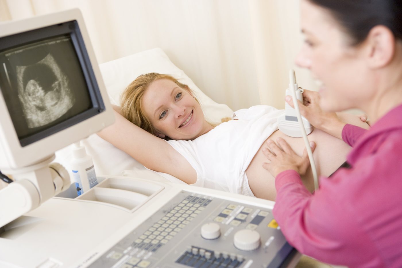
In early pregnancy, it’s best not to urinate before scans if possible, since a full bladder pushes up your womb, the ultrasound echoes will be able to reach your womb more easily, and the picture will be better. If you have an empty bladder they will ask you to drink a few glasses of water beforehand. At the scans, you lie down and they put a cold unscented gel on your tummy (though some kind sonographers warm it up!) A little scanning device (a transducer) is gently passed over the skin on your tummy by the person doing the scan (the sonographer). Sometimes if the sonographer needs to get a clearer image they may press down on your bump or in at the side which can be slightly uncomfortable (especially with a full bladder). The transducer beams a high-frequency sound into your womb. The sound bounces back and makes a picture, which you can see on a screen, a little like the way bats and dolphins use sound to map their surroundings and echolocate. You can usually take home a photo of this image.
Your doctor or midwife may recommend an earlier scan as well, but only if there are any problems, such as a previous miscarriage or bleeding. This scan might take place between 8-10 weeks. Sometimes these early scans might be carried out as a vaginal scan, rather than through your abdomen. This method gives a clearer picture at this early stage of pregnancy. It won’t be uncomfortable and you don’t need to keep a full bladder for this type of scan. You can ask for a female sonographer if you prefer.
The timing is between 10-14 weeks.
This scan looks at:
At the same time as your dating scan, you can also choose to have a combined screening to check for abnormalities.
This includes:
Please click HERE for more information from the NHS about antenatal scans.
Together these results help to calculate a statistical result of your baby’s chances of having Down’s Syndrome (the most common chromosomal disorder). This result is not a diagnosis but instead gives you an idea of how likely your results means that your baby may have the condition. You may get a result like a 1 in 34,000 chance.
Based on this result and your age you may want to take a diagnostic test to find out whether your baby has this syndrome. Some people don’t want to do the test and that is your choice. You will be at higher risk if you are an older Mum.
If you miss your chance to have this screening test you can have the Quadruple Blood Test that screens for levels of hormones in the blood that are higher in mothers carrying a baby with Down’s Syndrome and Edwards Syndrome only.
In reality, this scan occurs between 18-21 weeks. The sonographer will quickly freeze images of your baby to measure:
to see if your baby is growing well and in proportion.
Your sonographer will also look at the structure of important organs such as the heart, brain, umbilical cord, spine, abdominal wall, stomach, kidneys, bladder, arms, legs, feet and hands. In addition, the sonographer will note the position of the placenta to check it’s not too close to the cervix. If it is this will be checked again nearer birth.
The sonographer will measure to see how much amniotic fluid surrounds your baby. The amniotic fluid is produced by the baby, too much or too little fluid can indicate foetal distress.
If your sonographer is concerned about growth or anatomy, she may carry out a Doppler which records the oxygenation levels of the heart, umbilical cord and brain arteries to make sure your baby’s circulation system is working well. If there are any concerns you will be informed of your results in a consultation and may have more scans or treatment.
At this scan, you can also usually find out (if you want to) whether the baby is a boy or a girl. However, a scan isn’t 100% certain. Whether you decide to know the sex of your baby is entirely up to you. Some parents like surprise element at birth where others feel if they know the sex it can help them develop a closer connection during pregnancy with their little boy or girl.
If the sonographer can’t see everything he or she needs to at this scan, you may be asked in for a further scan a few weeks later.
In your third trimester, you may be asked in for further scans if you have previously had a very small baby, or if your baby is very small, or if you are carrying twins or triplets, if you have high blood pressure or diabetes, or if they are concerned for any other reason. You are unlikely to need a full bladder for these later scans since your baby should be so much bigger.
If the sonographer sees something they are worried about, they will send you to a doctor within 24 hours, or a foetal medical specialist, usually within 3 days.
The doctor or specialist may then carry out further tests, such as a CVS or amniocentesis. Please see our article on diagnostic tests for more information on these tests.
If the scan or tests reveal serious problems, talk to the doctors at length to have all your questions and options explored. There are a lot of people on hand to support you through any decisions that need to be made, including midwives, obstetricians, paediatricians and whoever else may be relevant to your particular situation. Make sure you get all the advice you need before making any decisions.